
A Case Study of a Plantar Arch Bump
-

Mr MC is a 39 year old male who presents to the clinic with a chief complaint of a painful “bump” in the arch of his right foot. He first noticed the bump approximately six months prior to presentation, initially it was relatively small and only mildly tender to touch, He has no recollection of injury to the area, or any other possible causation. The bump has slowly gotten larger and feels like it is forming additional bumps nearby. There is no redness, warmth or change in color to the area. He has tried massaging the bump to reduce its size and padding his shoe to relieve pressure on it when he stands and walks. His wife was concerned that he was getting a cancerous tumor in his foot, and after consulting with “Dr Google” online, determined that he was experiencing Plantar Fibromatosis. He also admitted his father had long suffered from a trigger finger in his hand. Upon researching treatment options, he discovered a high recurrence rate following surgical excision of plantar fibromas, and therefore is requesting an attempt at more conservative measures to alleviate his discomfort and hopefully prevent further growth of the bumps.
The patient is 5’!0” tall, and weighs 200 lbs. He works as a foreman in a factory and is on his feet all day in work boots on a concrete floor. He is relatively sedentary outside of work, taking an occasional walk with his dog. He denies smoking and drinks 1-2 beers per day after work.
Examination of both lower extremities finding include normal pedal pulses and capillary filling time bilaterally. Mild varicosities are noted in both lower legs and ankles with minimal edema and no hemosiderin deposition present. Hair and nail growth is normal, skin tone and temperature is normal. The patient has a mildly cavus foot type, with a supinated stance position. Ankle dorsiflexion is 5 degrees with the knee extended and 10 degrees with the knee flexed bilaterally.
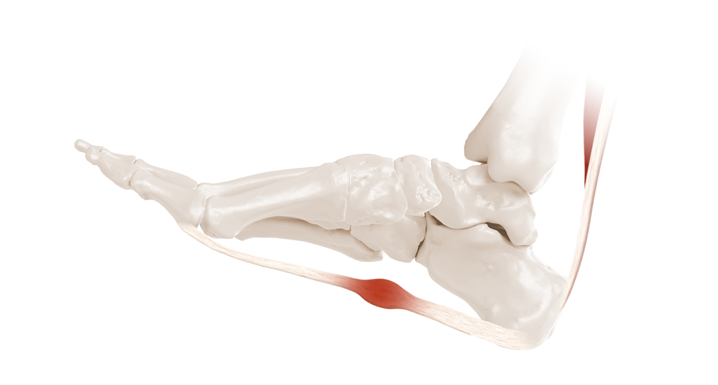
A multilobulated soft tissue mass is present in the midarch of the right foot along the medial band of the plantar fascia. It measures 1.5 cm in length and .75 cm in width and is tender to palpation. It blanches with digital pressure and is mobile beneath the skin. X-rays of the right foot were performed and show some soft tissue density without calcification in the area of chief concern.
Consultation regarding the treatment options and prognosis for plantar fibromatosis was conducted with the patient and his wife.They expressed a preference for the most conservative options with the understanding that plantar fibromatosis is somewhat unpredictable with the possibility of additional lesions arising. The use of a custom functional orthotic device was presented, and as a temporary test a low dye strapping with accommodative padding was applied to the right foot with the instruction of leaving it in place for 3-4 days, otherwise proceed with normal activities and return to the clinic in one week. The patient did return one week later reporting 90% reduction of the discomfort with the strapping and padding intact with some recurrence of the pain following its removal. The decision was then made to proceed with the manufacture of custom functional orthotics.
Discussion:
Common sense would dictate that using a soft, forgiving material to use in a case like this would be most appropriate. However, my experience has been that maintaining as much functional control of the foot with a more supportive device is more effective in the long run. A frame consisting of polypropylene or subortholene with very specific accommodative padding with poron or plastazote with a soft top cover of neoprene works quite well in many cases. An all EVA device is also a very good option. I believe it is imperative to maintain the medial arch as much as possible and shorten the distance between the proximal and distal ends of the plantar fascia to reduce longitudinal strain on the fascia. It is common for the size and location of the fibromas to change over the course of time so one should be prepared to adjust the devices periodically to allow for these changes as well as normal wear and tear. It is also of critical importance to mark the lesions very specifically on your scan or cast, as well as provide photos with measurement markings to allow the lab technicians to build the device as accurately as possible.