
Neuro Exam
-

In a previous edition of this column, we began an initial discussion of the documentation required to support the medical necessity for a drop foot brace. This week’s column will start a discussion of the proper lower extremity neurological examination and how it can support the need for an AFO.
As with all medical investigations, an essential medical history is necessary. Does the patient have an existing neurological diagnosis and are they under the active care of a neurologist, physiatrist, etc. for this condition? The patients’ various ADL limitations, use of ambulatory assistive devices and any fall history must be recorded in the medical record.
Some common examples of neurological diagnoses endemic to patients seen by podiatric physicians include but are not limited to those with a history of CVA or concussive syndrome (or post any head trauma), spinal stenosis, high fibular fracture effecting the common peroneal nerve, neuropathies (metabolic and post traumatic). If any are present, these should be well documented with date of onset, etiology etc. Previous MRI and other physician consultation reports including neurology, physiatry, radiology should all be documented and imported into your patient’s medical record.
The following essential neurological assessments, many courtesies of Dr. Richard Blake, can be used to meet the medical necessity requirements required by the LCD.
Observation: Look for signs of possible neurological involvement in gait and physical observation. The examiner should look for these signs: drop foot, steppage gait, Trendelenburg gait, antalgic gait, muscle atrophy, fasciculations, skin changes of shiny skin or loss of hair, and possible ulcerations.
The observation of any of these elements should be a driving force in having the examiner objectively dig deeper into further elements of a neurological exam. In many cases, these maneuvers should be repeated with the patient’s eyes open/closed and with and without resistance. It is also important to compare left vs. right. Documenting WNL (within normal limits) is NOT acceptable). These examination points include:
- Sensory examination: Defects in: Light Touch, sharp/dull discrimination, vibratory sense, proprioceptive, and protective sensation with 5.07 monofilament wire, temperature sensation (cold vs. heat perception).
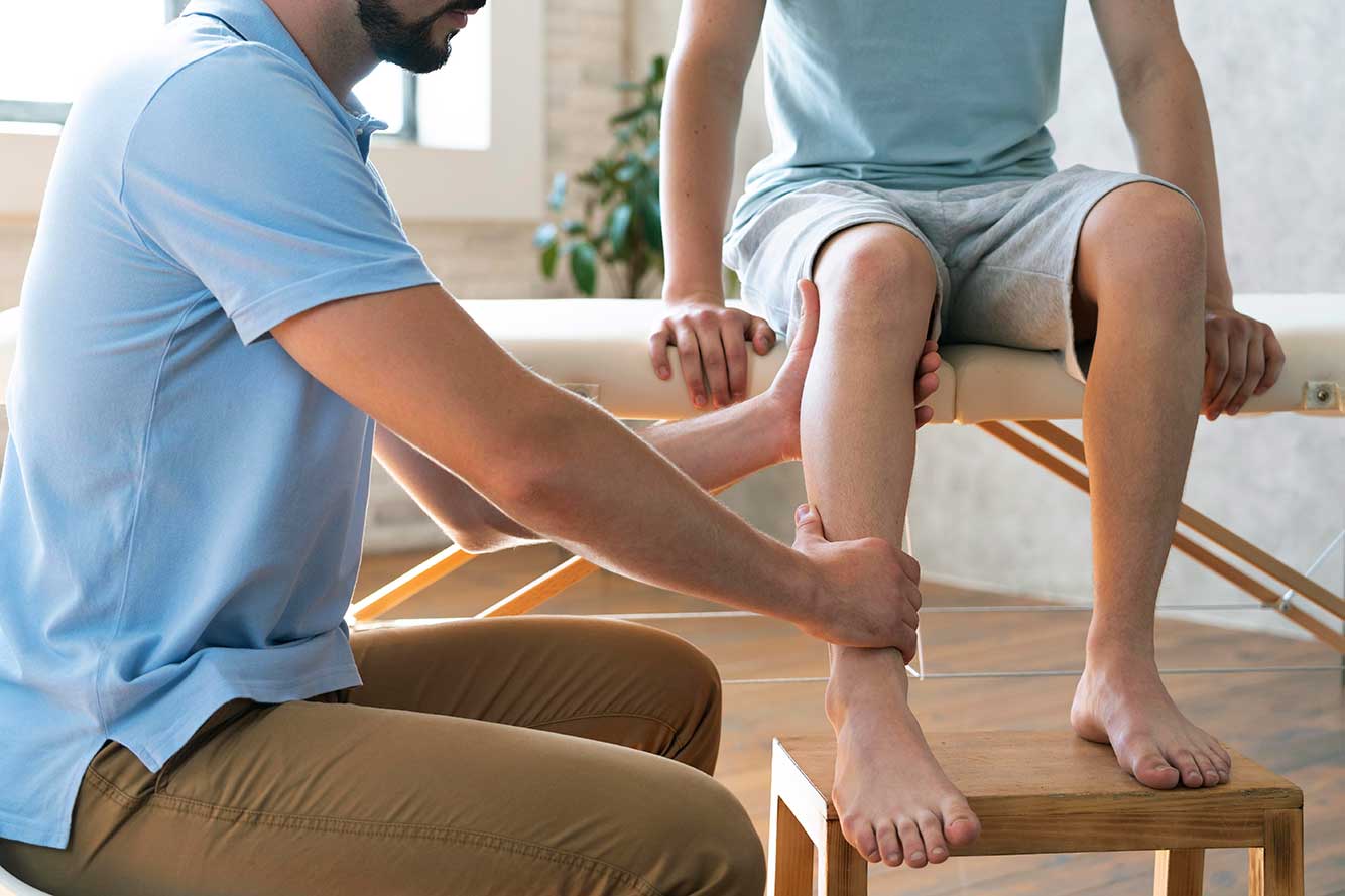
- Motor examination: Look for strength (motor tone) and fatigability. Especially test strength of ankle inversion/eversion, ankle dorsiflexion and plantarflexion, and hallux dorsiflexion. As necessary, compare these maneuvers side to side looking for asymmetry, both with and without resistance.
- Reflexes: Document Patellar and Achilles reflexes for absence, diminished, normal, brisk, or clonus responses.
- Neural Tension/Provocative Testing: These include slump test, straight leg testing, neural flossing with provocation, and Tinel’s sign.
- Coordination Testing: Common examples are heel to shin and rapid alternating heel taps.
- Especially for the elderly, quadriceps testing by having the patient rising from a seated position and sitting down with and without the assistance of chair arms should also be conducted. Changes from side to side are most often indicative of some neurological impairment.
- Tremors. Bilateral upper and lower extremities. Are they at rest, intention (just prior to movement) or limited to during motion?
- Range of Motion: Is range of motion met with resistance by the patient? Does it change with the patient’s eyes open vs. closed? Is there pain with ROM in one or more planes and along specific dermatomes?
- Balance Testing: Can the patient balance on one leg? If so, how long?
- Shove test: When the patient is standing, support their back to prevent them from falling and gently push them on their chest. Can they support themselves and not lose balance?
All these elements should be documented in an objective fashion. Once documented, a conclusionary statement with supportive rationale can be made. This conclusionary statement (e.g. diagnosis) and functional assessment on how the AFO will improve the patient’s gait can then be documented.
If this is a new diagnosis and the patient has not been seen by a neurologist or physiatrist a referral to one of these specialists may be in order. The more documentation one has in support of your diagnosis the better!
Some everyday clinical examples will follow in our next article in this series.