
First Ray Hypermobility
-

This week I am going to review this fine article on the topic of first ray hypermobility. It has long bothered me that anyone who overly-pronated was labeled as having a hypermobile first ray when that was not always the case. Hypomobile or normal first rays can be in patients who over-pronate. In Root biomechanics, normal first ray range of motion is 5-6 mm above the plane of the second metatarsal, and 5-6 mm below. Dr. Root defined more than this amount of 10 mm was hypermobility, and less than 8 mm was hypomobility. The author’s definition in the first paragraph was a little different.
In the second paragraph, the historical review is an important history with Dudley Morton (anatomist) and Dr. Lapidus. You can see that Dr. Root took Dr. Morton’s evaluation technique and fine-tuned it and placed numbers on it. Dr. Root stated that the examination must be done in subtalar neutral, and after thousands of examinations, stated the normal and abnormal values. Whereas I and Dr. Root use(d) First Ray Mobility Testing only in the sagittal plane, the article makes you aware of a more transverse plane version which has some validity if some of you reading want to research it.
To make sure you read this good review article, answer the following:
- Why do they propose that there are no Morton’s neuromas in the first intermetatarsal space?
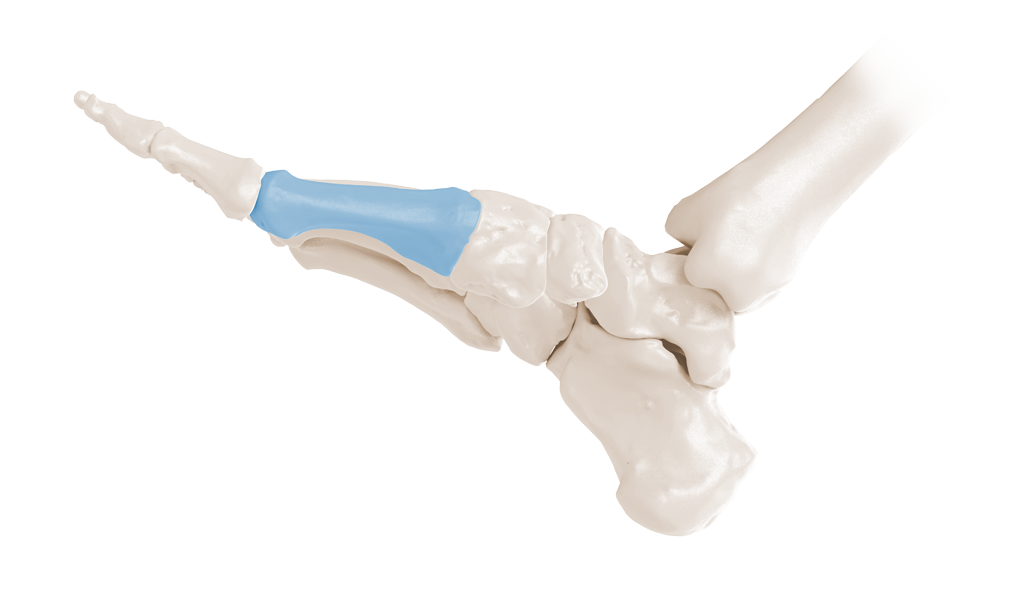
- What are all the joints involved in the First Ray?
- What two muscles are vitally important in First Ray stability?
- Does the Plantar Fascia play any role in First Ray stability?
- What does “Pronation of the FR” mean?
- What 2 planes do the authors feel are most important in accessing First Ray Hypermobility?
Dr. Root’s simple test from the 1970’s has withstood the test of time in my mind. It has been said he felt first ray hypermobility was occurring when the dorsal range was greater than the plantar range. I remember that hypermobility was when the measurements were over this 10-12 mm of normal range. If the first ray ROM was more dorsal then it was a dorsiflexed first ray (needing a Morton’s Extension on your Rx) and when the first ray ROM was more plantar then it was a Plantarflexed First Ray needing intrinsic balancing and a Reverse Morton’s aka Dancer’s Pad. It is also brought up in this article that the test should be done in ankle neutral to put tension on the plantar fascia, and therefore the test should be one of your patient Prone tests, not patient Supine tests. There are authors that say any dorsal excursion, even 1 mm, over the plane of the 2nd through 5th metatarsals is enough to call a first ray hypermobile, but that is in a small minority.
There are endless rulers to measure this excursion, but Dr. Root used his thumb thickness. I remember my class all walking around and measuring the thickness of our thumbs when slightly compressed from the dorsal to simulate the test. Any thumb thickness was fine. You just needed to know how to gauge the excursion. My thumb is 10 mm.
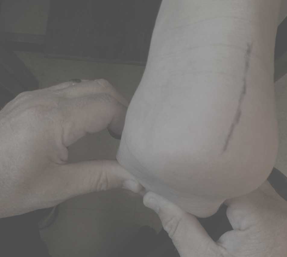
Here the patient is prone. I have stabilized the 2nd through 5th metatarsals. I attempt to get my thumbs parallel and apply a little pressure on the first metatarsal first upwards for dorsal excursion, and then plantar. This gives me my first ray ROM and I can classify the foot as hypermobile, hypomobile, dorsiflexed first ray, or Plantarflexed first ray. I know the thickness of my thumb and it is much less clumsy then the rulers out there.
There is more to the article if you care to read on. Of course, we need Dr. Doug Richie to weigh in on this discussion.