
Case Studies of the Use of Custom Functional Foot Orthotics #1
-

The next few weeks of posts will consist of hypothetical biomechanical cases intended to stimulate the thought process behind building an orthotic device. Biomechanical data will be provided, as well as subjective complaints and non-biomechanical objective findings. There is rarely one correct answer when deciding how to address these cases with an orthotic device alone, the reader may assume that other treatments may be used in addition to, or instead of an orthotic, however we will emphasize each case from a biomechanical or orthotic perspective. Recommendations may or may not be provided, some of the information provided may be very significant, while other information may be unimportant. The first case is a very common presentation, with a couple of wrinkles added, every practice should be familiar with this lady or someone like her!
Mrs. T.A. is a 52 year old female complaining of pain in the bottom of her heels, The pain has been present for at least three months, gradually worsening over that time. She does not recall any specific injury that might have caused the problem. She has tried wearing different shoes, putting heel pads in her shoes, stretching exercises, and over the counter medications, but nothing is helping.
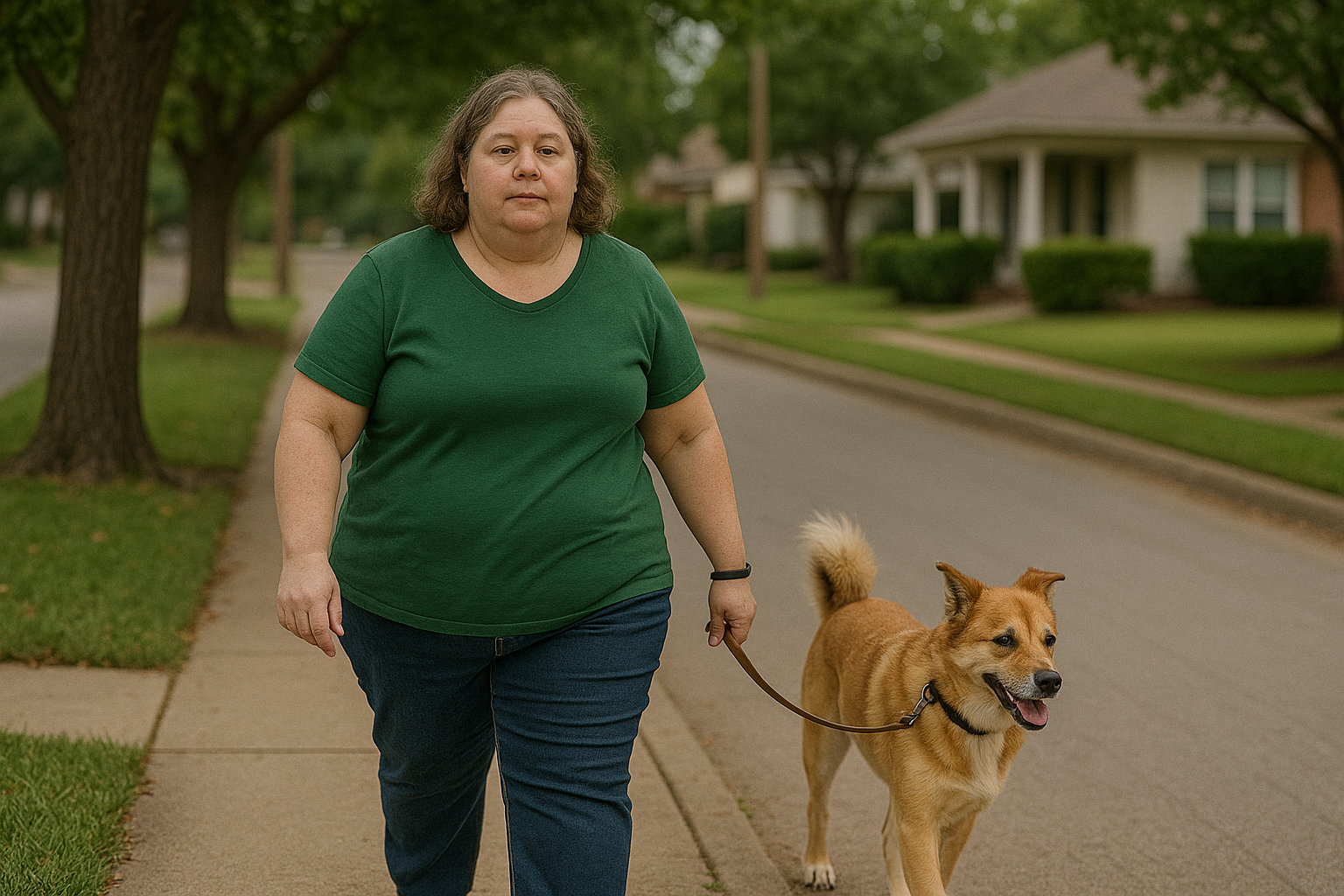
The patient is 5’ 2” tall and weighs 195 pounds. She works at a local supermarket as a cashier and stock person. She walks her dog 30-60 minutes, three times per week, and goes to Zumba classes one hour, twice a week. She is married, with two adult children who no longer live at home. She recently had a physical exam with her primary care physician, was told she had mild hypertension, but was otherwise healthy and instructed to modify her diet and exercise more to lose weight. She describes her heel pain as “dull and throbbing” with occasional episodes of sharper needle-like pain. The pain is worse while standing and working the check out line at the supermarket, not as pronounced when moving around the store stocking shelves. She says she has minimal pain after rest and in the morning which is lessened by wearing slippers at home rather than going barefoot. She wears non-slip sole oxford shoes at work, walking shoes for casual wear and walking her dog, and lightweight cross training shoes for Zumba. She rarely wears dress shoes.

Circulatory, neurological and dermatological exam findings of both lower extremities are found to be within normal limits and non-contributory.There is tenderness to palpation of both heels, mostly under the body of the Calcaneus, and with medial lateral compression, equal bilaterally. Minimal tenderness along the plantar fascia, no tenderness in either sinus tarsi, no palpable thickening or defect of the plantar fascia. No pain elicited with maximal passive ankle dorsiflexion. There are absent signs of acute inflammation. Weight bearing x-rays of both feet are negative for plantar Calcaneal spurs, with mild pronatory changes at the subtalar and midtarsal joints, negative for fracture, mildly elevated first metatarsal with mild joint space narrowing and spurring of the first metatarsal head.
Limited ankle dorsiflexion of 5 degrees is noted bilateral with knee extension, 10 degrees with the knees flexed. Her gait is antalgic, with symmetry noted in shoulder height, arm swing, hip drop, and knee position. She shows early heel off, moderate Calcaneal eversion in midstance and significant lowering of the medial arches,
We have decided to manufacture custom foot orthotics for the patient. Biomechanical exam findings include absent leg length discrepancy, genu valgum bilateral, 20 degrees external Tibial torsion bilateral, a bilateral neutral Calcaneal stance position of 2 degree inverted, a resting Calcaneal stance position of six degrees everted. Normal subtalar and midtarsal joint range of motion, moderate Navicular drop, 1st ray hypermobility, and forefoot supinatus of six degrees bilateral.
The patient is experiencing bilateral heel pain, without the classic signs of plantar fasciitis. What findings do we emphasize over others when creating our orthotic build? My standard practice when designing an orthotic is to break down the device into three main components, starting in the rearfoot, then midfoot and finally the forefoot and extension. As stated above, there is no single correct answer here, it is up to each practitioner to create their own decision making algorithm regarding posting, frame, and cover materials, pads and cushions and other modifications. What do you think?